Dermoscopy is a non-invasive diagnostic technique that has revolutionized the way dermatologists examine skin lesions. By utilizing specialized magnification tools, skin specialists can visualize structures beneath the surface of the skin that are invisible to the naked eye. This process of Dermoscopy Mole Evaluation in Abu Dhabi is a standard procedure for assessing skin health and monitoring changes in pigmented lesions. Understanding the necessity of this evaluation is the first step in proactive skin care.
- Dermoscopy uses specialized optics to visualize subsurface skin structures.
- It serves as a critical, non-invasive diagnostic tool for dermatologists.
- The technique allows for the early detection and monitoring of skin lesions.
- It is essential for differentiating between benign and suspicious moles.
What is Dermoscopy and How Does It Work?
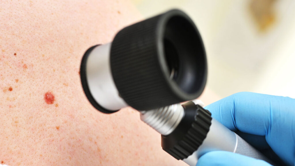
Dermoscopy, often referred to as dermatoscopy or epiluminescence microscopy, is a procedure that bridges the gap between clinical examination and surgical biopsy. When a mole appears suspicious, a dermatologist uses a dermatoscope—a handheld device combining a high-quality magnifying lens with a specialized lighting system. This setup eliminates surface light reflection, allowing the practitioner to peer into the epidermis and the upper layers of the dermis.
- The dermatoscope acts as a high-powered magnifying glass with polarized or non-polarized light.
- It eliminates skin surface glare, revealing underlying pigment patterns and vascular structures.
- This visual data helps dermatologists identify specific morphological features of a mole.
- The process provides a more accurate assessment than clinical inspection alone.
Identifying When a Mole Evaluation Is Necessary
The primary goal of monitoring skin health is early intervention. While most moles are benign, certain physical changes necessitate a professional evaluation. Using the “ABCDE” rule—Asymmetry, Border irregularity, Color variegation, Diameter enlargement, and Evolving shape or symptoms—is the standard method for identifying moles that require closer inspection. If a patient observes any of these changes, a thorough examination is warranted.
- The ABCDE rule provides a framework for self-monitoring skin health.
- Asymmetry or irregular borders are key indicators that a mole should be checked.
- Rapid changes in size, shape, or color are critical warning signs.
- Dermoscopy allows specialists to see if these physical changes correlate with concerning underlying structures.
The Role of Dermoscopy in Early Detection
The primary value of this diagnostic approach is its ability to detect structural changes long before they become visible to the naked eye. In many cases, a mole may look unremarkable under standard lighting but reveal irregular patterns under dermoscopic examination. This early detection is vital for ensuring that any necessary action can be taken promptly and effectively.
- Dermoscopy detects architectural irregularities invisible to the naked eye.
- It increases the diagnostic confidence of dermatology professionals.
- The technique aids in identifying markers that distinguish benign growths from others.
- Regular monitoring provides a historical baseline for an individual’s skin health.
Understanding the Evaluation Process
When a patient undergoes a professional evaluation, the process is straightforward and painless. After a thorough review of the patient’s skin history, the dermatologist will systematically examine specific lesions of interest. The use of the dermatoscope allows the specialist to map the mole’s features, including the distribution of melanin and the arrangement of skin lines. This mapping helps in tracking the stability or progression of the mole over time.
- The process begins with a review of medical and sun-exposure history.
- The specialist systematically examines each mole of concern.
- Dermoscopic images are often compared to previous records for changes.
- The procedure is entirely non-invasive and requires no recovery time.
Why Proactive Monitoring Matters
Skin health is dynamic; moles can change over time due to hormonal fluctuations, sun exposure, and natural aging processes. By integrating routine evaluations into a healthcare regimen, individuals can maintain a clear understanding of their skin’s unique topography. This proactive approach takes the guesswork out of skin health and provides peace of mind through documented observations.
- Moles are dynamic and can change throughout a person’s life.
- Environmental factors like sun exposure heavily influence skin lesion development.
- Routine evaluations create a longitudinal record of skin health.
- Proactive monitoring reduces anxiety regarding the status of unusual-looking moles.
Frequently Asked Questions
Does dermoscopy cause any discomfort to the skin?
No, the procedure is entirely non-invasive. The dermatoscope simply touches the skin or hovers slightly above it using light to visualize the subsurface, causing no pain or damage.
How often should I have my moles evaluated?
The frequency of evaluations depends on your personal history, skin type, and the number of moles you have. It is generally recommended to discuss a personalized screening schedule with a professional during your initial consultation.
Can dermoscopy identify all skin concerns?
Dermoscopy is a highly effective tool for evaluating pigmented and non-pigmented lesions, but it is one part of a comprehensive skin health assessment. A dermatologist will combine these findings with your medical history and clinical presentation to reach a conclusion.
What should I do if I notice a mole changing?
If you notice any changes in the size, shape, or color of a mole, you should schedule an appointment for a professional assessment. Even if the change turns out to be minor, having it evaluated by a specialist ensures that you are staying ahead of your skin health.